fMRI vs MRI: What’s the Difference and Why It Matters

If an MRI is like a crystal-clear photo of your brain, an fMRI is the video where you can actually see what’s happening behind the scenes. Both scans use the same powerful magnet, but they’re built to answer totally different questions: One shows the brain’s structure, the other shows its activity in motion. Understanding that difference can make the whole idea of “getting a brain scan” feel a lot less mysterious (and a lot less intimidating).
If you’ve ever tried to make sense of the different brain scan options, you know how quickly it can get confusing. Thankfully, the core difference between fMRI vs. MRI is easy to grasp: An MRI shows what the brain looks like. An fMRI shows what the brain is doing.
MRI—aka magnetic resonance imaging—is essentially a detailed snapshot of the brain’s structure. An fMRI (functional MRI) uses the same magnet but adds a layer of motion so you can watch brain function (like blood flow) in 3D.
In this guide, we’ll walk through what each scan does, how they’re used, what they can (and can’t) tell you, and what to expect if your clinician recommends one.
What is an MRI?
An MRI (sometimes called a structural MRI) is a noninvasive scan that gives your doctor a clear, detailed picture of what’s happening inside your body. Think of it as a high-quality snapshot: sharp, precise, and especially good at showing the brain’s physical structure. Clinicians often use MRI to look for things like injury, inflammation, stroke-related changes, or natural shifts that come with aging—all of which can help explain symptoms or rule out other medical causes.
Here’s how it works, what it can show, and why it’s usually the first stop when someone needs a closer look at the brain.
How MRI works
At its core, an MRI is simply a giant magnet. “An MRI machine is a giant magnet so powerful it uses superconductors cooled by liquid helium that let us look at the different tissues in the body without using radiation,” says David Carreon, MD, a psychiatrist and CEO of Acacia Clinics.
The magnet lines up the hydrogen atoms in your body, radio waves gently nudge them, and as they settle back into place, the scanner detects the signals they release. A computer converts those signals into crisp, slice-by-slice pictures of your brain’s structure.
Here’s what that looks like in practice:
- The machine makes loud tapping and knocking sounds as it switches magnetic fields.
- You lie still in the machine so the computer can knit all the signals together into sharp MRI images of the brain.
- Sometimes you’ll get contrast dye through an IV to help highlight certain tissues or blood vessels (think of it like adding a highlighter to the inside of your body so specific areas stand out more clearly).
- Most scans take about 20–45 minutes.
What MRI shows
Because the images are so sharp, MRI can help identify changes or abnormalities that might explain certain symptoms. For instance, it can reveal:
- Tumors or mass-like growths
- Signs of stroke or reduced blood flow
- Inflammation or infection
- Multiple sclerosis lesions
- Traumatic brain injury
- Age-related atrophy or shrinkage
- Congenital (from birth) differences in brain structure
MRI is also used to image many other parts of the body—joints, the spine, organs—but in a mental health or neurology context, its main job is to give a clear picture of the brain’s anatomy, according to Carreon.
When MRI is used
Common reasons for a brain MRI include:
- New, severe, or persistent headaches
- Seizures or suspected epilepsy
- Memory changes or cognitive concerns
- Sudden neurological symptoms like weakness, numbness, or dizziness
- Symptoms that could be related to stroke
- Unexplained changes in mood or behavior where a structural cause needs to be ruled out
Clinicians order MRI scans when they want to rule out or better understand a physical issue that could be contributing to symptoms. Because it’s safe, noninvasive, and extremely detailed, MRI is often the first imaging tool used in both neurology and psychiatry.
Connect with our Clinicians


What is an fMRI?
Instead of showing what your brain looks like, an fMRI shows what your brain is doing by tracking changes in blood flow over time.
How fMRI works
Functional MRI uses the same giant magnet as a standard MRI, but “but you can ‘tune’ the MRI to look for blood as it flows through the brain,” says Carreon. “If you analyze this 3D video, you can get an idea of the function of the brain.”
Instead of focusing on tissue structure, it measures the blood-oxygen-level dependent (BOLD) signal, which is basically a way of seeing where fresh, oxygen-rich blood flows when different parts of your brain “switch on.” The result is like watching a heat map of your brain in action.
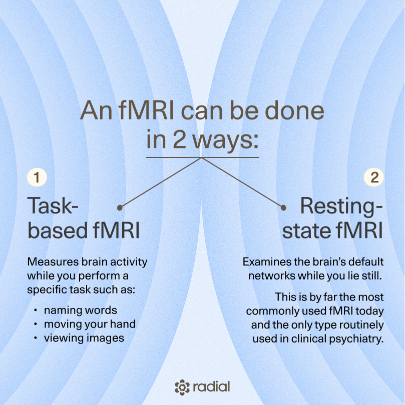
This can be done in two ways:
- Task-based fMRI, which is a scan for brain activity while you do something specific (like naming words, moving your hand, or looking at images)
- Resting-state fMRI, which looks at the brain’s default networks when you’re simply lying still. This is, overwhelmingly, the most common type of fMRI used today, and the only kind used in routine clinical care in psychiatry for treatments like SAINT TMS targeting.
Functional MRIs are not to be confused with structural MRIs, which are standard MRIs designed to show images of the brain’s anatomy. It’s sometimes the case that physicians will order both at once, so that they can accurately target their treatments based on both structure and function.
What fMRI shows
An fMRI of your brain shows how it’s behaving in real time—which areas are working, how strongly they activate, and how different regions talk to each other. Clinicians and researchers use fMRI to get a better sense of things like:
- Which parts of the brain support specific tasks (like speaking, moving, remembering, or processing emotions)
- How brain networks reorganize after an injury or stroke
- How activity patterns may differ in certain psychiatric or neurological conditions
- Whether certain brain circuits might be good targets for treatments like neurosurgery or TMS
When fMRI is used
fMRI has expanded far beyond research labs. Today, it’s used in a wide range of clinical and scientific settings, including:
- Neurosurgery planning: It helps surgeons identify important areas like speech or movement so they can avoid those regions during an operation.
- Stroke recovery: It shows how the brain reorganizes and heals over time.
- Chronic pain research: Scientists use it to study how pain-related circuits activate and change in different conditions.
- Psychiatry: “The most exciting new use of these technologies are in models of treatment that use fMRI to find targets to deliver treatments,” says Carreon. “Targets exist for conditions ranging from depression, to PTSD, to pain. They use magnetic stimulation to induce plasticity and help patients recover.” This includes precision approaches like SAINT® TMS, where fMRI helps map the specific brain circuits involved in treatment-resistant depression and guide stimulation directly to the right spot.
- Scientific research: fMRI continues to be a cornerstone tool for studying everything from memory and emotion to language and learning.
fMRI has a lot of uses: However, it can’t diagnose a mental illness by itself. It also can’t read your mind or work as a lie detector (no matter what sci-fi movies or wellness marketing might promise).
fMRI vs. MRI: Key differences
If MRI is the photo and fMRI is the video, the biggest difference between the two comes down to what they measure and why they’re used. Here’s a closer look at structural MRI vs. functional MRI, and how they differ in everyday clinical practice:
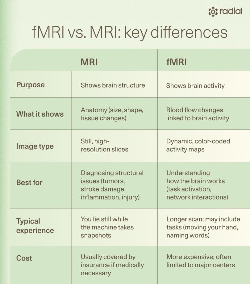
- What they measure: MRI captures detailed pictures of brain anatomy, while fMRI tracks changes in blood flow to reveal brain activity. It’s common to get a structural MRI alone, but fMRI almost always includes a structural MRI image, too.
- What the images look like: MRI produces still, high-resolution images. fMRI produces dynamic maps that change over time as different areas “switch on” as well as the relationship between different brain circuits as they work together.
- Typical use: MRI is commonly used to diagnose structural issues like tumors, stroke damage, inflammation, or injury. fMRI is used when clinicians need to understand how the brain is functioning, often for neurosurgery planning, research, or treatments that target specific brain circuits.
- Duration and experience: Both scans happen in the same machine, but fMRI tends to take longer because it captures continuous activity. You may also be asked to perform tasks (like moving your hand or naming words) during an fMRI, whereas you lay still during an MRI.
- Cost and availability: MRI is widely available and often covered by insurance. fMRI is less common, more expensive, and often limited to large medical centers or research hospitals.
Like any tool, each scan has its strengths and limitations: MRI is versatile, widely available, noninvasive, and excellent for spotting structural problems. However, it can’t measure brain function, and some people find the noise or enclosed space uncomfortable.
On the other hand, fMRI benefits include getting a dynamic view of how the brain works and how different regions interact, which can be extremely useful for planning surgery or targeting treatments like TMS. Still, there are fMRI disadvantages: It’s expensive, harder to access, and very sensitive to motion—even small movements can blur the results.
When doctors choose one over the other
Clinicians generally choose fMRI vs MRI based on the question they’re trying to answer:
- MRI is used when the priority is understanding the brain’s structure. The question here is often something like: Is there damage, inflammation, or another physical issue that could explain these symptoms?
- fMRI is used when the goal is to understand function. The guiding question becomes: How is this person’s brain working, and which circuits are involved?
In some cases, the two scans complement each other: MRI gives the map, and fMRI shows the traffic patterns.
What to expect during an MRI or fMRI
Whether you’re getting an MRI or an fMRI scan, the experience is more similar than different. Both happen in the same type of machine—a large, tube-shaped magnet—and are completely noninvasive. The biggest distinctions are how long the scan lasts and whether you’ll be asked to do anything during it.
Before the scan, you’ll change into a gown or remove anything metal (like jewelry, hairpins, certain clothing). A technician will make sure you’re comfortable and answer any questions you have. If you’re getting contrast dye, you’ll receive a small IV beforehand.
Once you’re lying on the table, the technician slides you into the machine and gives you earplugs to minimize the loud tapping sounds (which, by the way, are totally normal). Neither scan hurts, and you won’t feel the magnetic field or radio waves. You’ll also be able to communicate with the tech at any time through a speaker system.
Here’s how the experience differs:
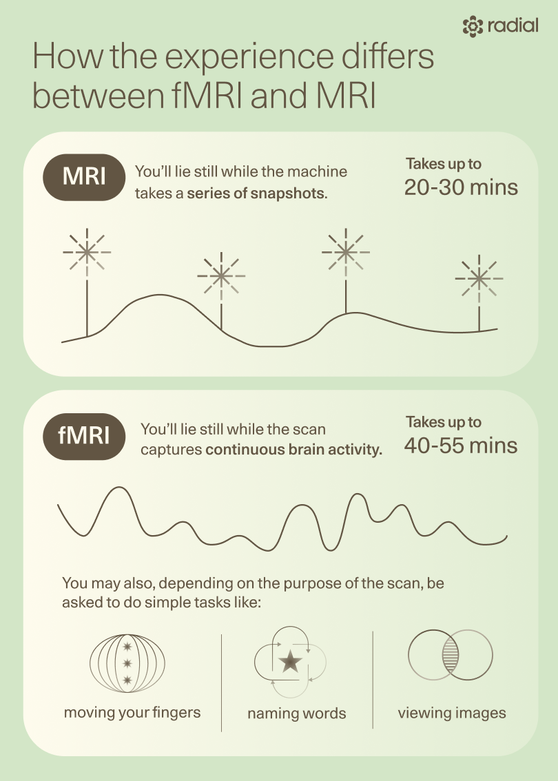
- MRI: You’ll lie still while the machine takes a series of snapshots. This usually takes about 20–30 minutes.
- fMRI: You’ll lie still as well, but the scan takes longer (20-55 minutes) because it’s capturing continuous activity. You may also be asked to perform simple tasks (like moving your fingers, naming words, or looking at images) depending on the purpose of the scan. If you’re feeling a little nervous, that’s also completely normal. Here are a few ways to make the scan feel a lot easier:
- Let your technician know if you’re anxious—they’re great at helping people through it.
- Ask for a blanket or extra padding to help you stay still and comfortable.
- Close your eyes before going into the machine if you’re prone to claustrophobia.
- Focus on slow, steady breathing to stay relaxed.
- If you’re doing an fMRI task, don’t worry about “getting it right”—the goal is simply to follow instructions at your own pace.
The future of brain imaging
Functional brain imaging is moving quickly, and many of the tools that once lived only in research labs are starting to influence real-world care. It’s still early, but the direction is clear: the future of brain imaging includes more personalized maps of how our brains work and more targeted treatments based on those maps, according to Carreon. For people who’ve struggled with trial-and-error treatments, this kind of progress offers real hope.
One major area of growth is precision psychiatry, where clinicians use personalized fMRI maps to see how someone’s brain circuits communicate. Instead of guessing which areas might be involved in depression, PTSD, or chronic pain, fMRI can help show the exact networks at play.
At Radial, clinicians already use this approach, combining MRI and fMRI imaging to guide highly targeted treatments like SAINT® TMS. And as imaging continues to improve, more research can identify new targets for precision fMRI-guided treatments, and can make treatment cheaper and more accessible
Researchers are also studying real-time fMRI neurofeedback, where people can watch their own brain activity shift and learn to influence it. Early studies are interesting—especially for depression, anxiety, and mood symptoms—but this type of neurofeedback is still very much in the experimental stage and requires equipment you won’t find in a typical clinic.
Another fast-moving area of research is neuroplasticity research, which looks at how the brain heals and rewires itself after injury or illness. fMRI helps scientists see those changes as they happen, offering clues that could shape future rehab and mental health treatments.
Finally, you may also hear about brain–computer interfaces, including headline-grabbing projects like Neuralink. These tools aim to help people with paralysis or cerebral palsy by translating brain activity into digital signals. It’s a fascinating field, but it’s still early, often overhyped, and usually relies on tools other than fMRI.
Relief within reach
Care covered by your insurance
Radial provides advanced mental health treatment, covered by the insurance you already use.


The bottom line
MRI and fMRI each offer a different window into your brain: MRI shows its structure, and fMRI shows its activity in real time. Your clinician may recommend one or both depending on what they’re trying to learn. These scans can’t diagnose mental illness, but they can give clarity, rule out medical concerns, and help guide treatments that target the brain circuits involved in your symptoms.
As imaging tools continue to advance, they’re opening the door to mental health care that feels more personalized, more effective, and more hopeful. At Radial, clinicians use both structural MRI and fMRI together to guide SAINT® TMS targeting and support clinical research for PTSD, bipolar depression, OCD, and treatment-resistant depression recovery. This integrated approach helps move mental health treatment away from trial and error and toward truly precision-guided care.
Key takeaways
- MRI and fMRI are not interchangeable. MRI produces images that show the brain’s structure; fMRI produces videos that show its activity.
- Both scans use the same machine, but fMRI takes longer and may involve simple tasks, while an MRI just requires you to lie still.
- MRI is great for spotting physical issues like stroke damage, inflammation, tumors, or injury. fMRI adds another layer by showing how brain regions “talk” to each other and helping guide circuit-focused treatments like TMS and newer precision approaches.
- Brain imaging is moving toward more personalized care, offering new hope for people who haven’t responded to traditional treatments.
Frequently asked questions (FAQs)
What is the difference between MRI and fMRI?
An MRI shows the brain’s structure: the size, shape, and health of different regions. An fMRI shows the brain’s activity by tracking blood flow changes over time. Think of MRI as a photo and fMRI as a video. Depending on what your clinician is looking for, you may need one or both to get the full picture.
Is fMRI invasive?
No, fMRI is completely noninvasive. It uses the same type of magnetic scanner as a standard MRI—no needles, no radiation, and no pain. The only difference is that you may be asked to do simple tasks (like moving your fingers or naming words) while the fMRI machine tracks how different parts of your brain respond.
How long does an fMRI take?
Most fMRI brain scans take between 20 and 55 minutes. They run longer than standard MRIs because they capture continuous activity over time. If you’re doing a task-based fMRI, you’ll spend part of that time following simple instructions while the machine measures how your brain reacts.
Editorial Standards
At Radial, we believe better health starts with trusted information. Our mission is to empower readers with accurate, accessible, and compassionate content rooted in evidence-based research and reviewed by qualified medical professionals. We’re committed to ensuring the quality and trustworthiness of our content and editorial process–and providing information that is up-to-date, accurate, and relies on evidence-based research and peer-reviewed journals. Learn more about our editorial process.
Let's connect
Get started with finding the right treatment for you or someone you care about
Get startedExplore more posts